


What do a woman suffering from Psoriasis, a man dying after acute rejection of kidney transplant, and a child suffering from Pulmonary Tuberculosis due to susceptibility to mycobacteria infection all have in common? These situations and conditions all involve the Human Leukocyte Antigen (HLA) System. Our body’s immune system uses HLA to distinguish between our own and foreign cells, and between healthy and infected cells. Think of HLA as a person’s signature. HLA proteins are encoded by a cluster of Major Histocompatibility Complex (MHC) genes located on chromosome 6 and are highly variable. While HLA is often associated with bone marrow and organ transplant, HLA typing is also used to identify disease-related biomarkers and has a role in vaccine development.
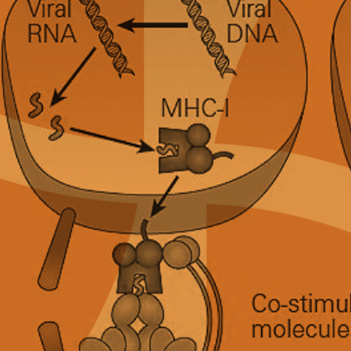
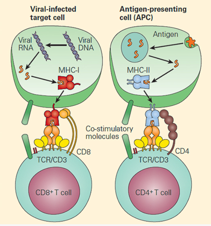
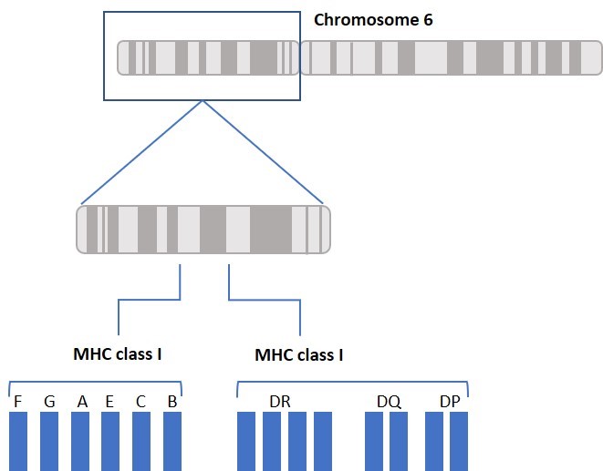
The main function of HLA is to educate our immune cells to distinguish between self and non-self. HLA molecules also play a role in initiating and coordinating immune response by binding and presenting antigens. HLA genes that involve the immune system belong to two major groups: MHC class I and MHC class II. Class I molecules are encoded by HLA-A, HLA-B, and HLA-C genes/loci and present intracellular antigens originating from viruses or tumors to cytotoxic CD8+ T cells. Class I HLA-G proteins play a role in immune tolerance (like maintaining fetal-maternal tolerance1), and Class I HLA-E proteins react with natural killer (NK) cells. Activation or inhibition of NK cells is achieved through crosstalk between killer immunoglobulin-like receptors (KIRs) on NK cells and HLA. (More about KIRs to come in a future blog.) Class II molecules are encoded by HLA-DP, HLA-DQ, HLA-DR, HLA-DM, and HLA-DO, and are mainly found on antigen presenting cells and present extracellular antigens to CD4+ T helper cells. MHC class III genes/loci do not encode HLA molecules. The MHC class III region encodes complement components, tumor necrosis factors (TNFs), and others (Figure 1).
([Bellanti, JA (Ed). Immunology IV: Clinical Applications in Health and Disease. I Care Press, Bethesda, MD, 2012)
Everyone’s HLA complex consists of unique variants of class I and class II genes, also called alleles. The string of alleles is referred to as a haplotype. We inherit one haplotype from each parent, and the two form each person’s HLA genotype. HLA typing could be done based on serology, which detects anti-HLA antibodies, or by DNA sequencing, which provides specific allele information. We use high resolution DNA sequencing to provide HLA genotypes on all of our cell products. In allogeneic organ or cell transplantation, it is much easier to find an HLA matched donor in the patient’s family. For siblings, the probability of having 2 identical haplotypes is 25%, the probability of having one identical haplotype is 50%, and the probability of having two different haplotypes is 25%. The probability of finding an unrelated matched donor depends on ethnicity and ranges between 16% (for black people of South or Central American descent) and 75% (for white people of European descent). 4
When it comes to organ or bone marrow transplantation, an optimal unrelated donor will require at least 8/8 allele match with the recipient at the HLA-A, HLA-B, HLA-C, and HLA-DRB1 loci. Various studies revealed that these specific genes are associated with graft rejection and patient survival compared to the rest of the HLA genes.2,3 A 7/8 allele match (i.e., one mismatch) reduces the 5-year overall survival rate by about 8%. Matching requirements for cord blood are less stringent (6/6 matching of HLA-A and HLA-B and HLA-DRB1). This is due to the increased proliferative capability of cord blood hematopoietic stem cells and the immature nature of cord blood immune cells, which lowers the risk for rejection and graft-versus-host (GvHD) disease.5-7 GvHD occurs when the donor’s cells attack the recipient’s healthy cells. This response is due to cross-reactivity in an HLA mismatched transplantation.
Just as in the case of organ and tissue transplantation, it is important to consider HLA matching when developing allogeneic cell therapies, such as mesenchymal stem cell (MSC) and immune cell therapies. Although MSCs express low levels of HLA I and low/no HLA II and are sometimes considered immune-privileged and better tolerated, immune reactions should still be considered. It has been reported that bone marrow-derived allogeneic mismatched MSCs elicited an immune reaction in vivo. Therefore, in order to develop safe and effective treatments, we must consider the immunological interactions between donor and recipient cells.8,9 All of OrganaBio’s cell products are HLA typed in order to better inform researchers and to ensure safe and effective cell therapies.
Some specific HLA types are associated with protective immunity versus susceptibility to infectious diseases. HLA genes have evolved through natural selection by selective pressure from infectious pathogens, with malaria being one of them. HIV vaccine development relies on specific HLA types targeted by vaccine peptides which induce protective immunity. HLA typing has also led to some improvement in the diagnosis of celiac disease and type 1 diabetes. Additionally, several autoimmune diseases have been correlated with specific HLA-B27 alleles. More on this in a future post.
Allele – A variant form of a gene, or locus
Haplotype– The combination of alleles from two or more loci located on the same chromosome
Genotype – The combination of two haplotypes; one from each parent, inherited by an individual
References
- Ferreira LMR, Meissner TB, Tilburgs T, Strominger JL. HLA-G: at the interface of maternal-fetal tolerance. Trends Immunol. (2017) 38:272–86.
- Lee SJ, Klein J, Haagenson M, et al. High-resolution donor-recipient HLA matching contributes to the success of unrelated donor marrow transplantation.
- Horan J, Wang T, Haagenson M, et al. Evaluation of HLA matching in unrelated hematopoietic stem cell transplantation for nonmalignant disorders. Blood. 2012; 120:2918–24.
- Gragert L, Eapen M, Williams E, et al. HLA match likelihoods for hematopoietic stem-cell grafts in the U.S. registry. N Engl J Med. 2014;371(4):339-348.
- Atsuta Y, Suzuki R, Nagamura-Inoue T, et al. Disease-specific analyses of unrelated cord blood transplantation compared with unrelated bone marrow transplantation in adult patients with acute leukemia. Blood. 2009; 113:1631–8.
- Gluckman E, Rocha V, Arcese W, et al. Factors associated with outcomes of unrelated cord blood transplant: guidelines for donor choice. Exp Hematol. 2004; 32:397–407.
- Dessels C, Alessandrini M, Pepper MS. Factors Influencing the Umbilical Cord Blood Stem Cell Industry: An Evolving Treatment Landscape. Stem Cells Transl Med. 2018;7(9):643-650. doi:10.1002/sctm.17-0244
- Isakova I.A., Lanclos C., Bruhn J., Kuroda M.J., Baker K.C., Krishnappa V., Phinney D.G. Allo-reactivity of mesenchymal stem cells in rhesus macaques is dose and haplotype dependent and limits durable cell engraftment in vivo. PLoS ONE. 2014;9:e87238.
- Pezzanite L.M., Fortier L.A., Antczak D.F., Cassano J.M., Brosnahan M.M., Miller D., Schnabel L.V. Equine allogeneic bone marrow-derived mesenchymal stromal cells elicit antibody responses in vivo. Stem Cell Res. Ther. 2015;6:1–11.
Donor selection
From HLA background to an actual cohort
Knowing the biology is one thing; finding enough donors who carry the haplotype is the part that stalls studies. OrganaBio publishes HLA data at the lot level and can go back to the same donors for repeat collections.
HLA and MHC: the same thing, named twice
The major histocompatibility complex is the general term, used across species. HLA, human leukocyte antigen, is what that complex is called in humans. Mouse immunologists say H-2. Same system, different species, different label. When a paper uses MHC and a certificate of analysis uses HLA, they are not describing two different things.
The name is a historical accident worth knowing, because it explains the confusion. These molecules were discovered as antigens on white blood cells that provoked antibody responses after transfusion and pregnancy, hence “leukocyte antigen”. Their actual job, presenting peptides to T cells, was worked out later. The name records how they were found rather than what they do.
Where the genes sit, and why that matters
The HLA region occupies roughly four megabases on the short arm of chromosome 6, at 6p21. It is one of the most gene-dense stretches of the human genome and by some distance the most polymorphic. Three sub-regions sit in a row: class I at the telomeric end, class III in the middle, class II toward the centromere.
Because these genes are packed close together, they are inherited as a linked block rather than shuffled independently. That single structural fact drives everything downstream: sibling match probabilities, the population-specific haplotype frequencies that make unrelated donor matching harder for some ancestries than others, and the linkage disequilibrium that makes certain allele combinations far more common than chance would predict.
What the molecules look like and how they work
| Class I | Class II | |
|---|---|---|
| Chain structure | One polymorphic heavy chain plus invariant β2-microglobulin | Two polymorphic chains, α and β |
| Peptide groove | Closed at both ends | Open at both ends |
| Peptide length held | Short, typically 8 to 10 residues | Longer and more variable, often 13 to 25 |
| Peptides come from | Inside the cell, degraded by the proteasome | Outside the cell, taken up and processed in endosomes |
| Presented to | CD8+ T cells | CD4+ T cells |
| Expressed on | Every nucleated cell | Antigen-presenting cells, and inducible elsewhere |
The groove geometry explains the length difference and it is not trivia. A closed groove clamps a short peptide at both ends. An open groove lets a longer one hang out either side. This is why class I and class II epitope prediction are different problems, and why an assay designed around one does not transfer cleanly to the other.
Class III sits physically between them and encodes complement components, tumour necrosis factor and heat shock proteins. It presents nothing. It is in the HLA region by address, not by function, which is a recurring source of confusion in exam questions and in supplier documentation alike.
MHC restriction, and why your T cells only work in your body
A T cell does not recognise a peptide floating free. It recognises a peptide held in a specific HLA molecule, and it recognises the two together as a single composite shape. This is MHC restriction. A T cell primed against a viral peptide presented on HLA-A*02:01 will not respond to that same peptide presented on a different allele.
Two consequences follow, and both are practical rather than academic. Epitope work is allele-specific, so a T cell assay without donor HLA type attached produces variance you cannot explain afterwards. And transplanted T cells encountering unfamiliar HLA react against it, which is the molecular origin of both graft rejection and graft-versus-host disease.
Polymorphism: the reason any of this is hard
Tens of thousands of HLA alleles have been catalogued, and the variation is concentrated precisely where it matters, in the residues lining the peptide-binding groove. That is not random drift. A population in which everyone presented the same narrow peptide repertoire would be a population a single pathogen could clear. Diversity here is a species-level immune strategy.
What protects the species complicates the clinic. It makes unrelated donor matching a probability exercise, it makes epitope prediction allele-dependent, and it makes “HLA-typed” a claim whose value depends entirely on the resolution behind it.
HLA and disease
Some of the strongest genotype-phenotype associations in human medicine sit in this region.
| Allele | Association | Why it is cited |
|---|---|---|
| HLA-B*27 | Ankylosing spondylitis | One of the earliest and strongest associations described |
| HLA-B*57:01 | Abacavir hypersensitivity | The reference example of HLA-guided drug safety |
| HLA-B*15:02 | Severe carbamazepine reactions in specific populations | Pharmacogenomic screening in practice |
| HLA-DQ2 and DQ8 | Coeliac disease | Near-necessary though not sufficient |
| HLA-DR3 and DR4 | Type 1 diabetes | Major contributor to genetic risk |
Association is not causation and none of these are diagnostic on their own. Their practical value is in stratifying research cohorts and, for the drug hypersensitivity alleles, in avoiding a foreseeable harm. For research programmes studying autoimmune disease, being able to select disease-state donor material by HLA genotype rather than by diagnosis alone is often the difference between a clean cohort and a noisy one.
HLA in cell therapy
Autologous programmes sidestep the problem: the patient’s own cells carry the patient’s own HLA. Allogeneic programmes cannot, and how they handle it defines the product.
Three broad strategies are in play. Match donor to recipient, which limits reach and needs a deep, well-characterised donor pool. Delete class I, usually by knocking out β2-microglobulin, which hides the cell from T cells but creates a new problem, because a cell with no class I is exactly what NK cells are built to kill. Or select an effector type that tolerates mismatch, which is a large part of why NK cells and gamma-delta T cells attract allogeneic interest.
The second strategy is worth sitting with, because it is where HLA and NK biology meet. Removing class I to evade T cells hands the cell straight to NK surveillance, which is why hypoimmunogenic designs increasingly retain HLA-E or HLA-G to keep the inhibitory NK signal alive. Understanding what NK cells do with that signal is covered in the NK cell guide.
For material with HLA type documented up front, OrganaBio’s donor characterisation programme runs across the catalogue, including leukopaks, cryopreserved PBMCs, pan T cells and NK cells. Donor selection can be scoped by HLA genotype subject to availability, and eligible donors can be scheduled for repeat collection.