


While Human Leukocyte Antigen (HLA) matching is often emphasized in the context of bone marrow and organ transplant, HLA typing is used to identify disease-related biomarkers and predict immune responses, including those that could lead to cell rejection and/or graft-versus-host disease (GvHD), which is why researchers are interested in HLA phenotyping and genotyping of cellular starting material. The major histocompatibility complex (MHC) region on chromosome 6, where the HLA genes are encoded, is one of the most complex and polymorphic regions in the human genome. Although there are more than 200 HLA genes, 6 are crucial for the distinction between self and non-self. HLA-A, HLA-B, and HLA-C genes/loci belong to class I, and HLA-DP, HLA-DQ, and HLA-DR belong to class II. More details about HLA basics can be found in our previous blog post.
HLA typing can be performed based on serology at the antigen level, which detects anti-HLA antibodies, or at the DNA level by sequencing, which provides specific allele information. Serology testing used to be the “gold standard” HLA typing, relying on histocompatibility assays which identify molecules that are actually expressed on the cells and are detected by monoclonal antibodies. It is quick, simple, and low cost test that established an HLA phenotype. However, serological HLA typing doesn’t account for sequence variation like some differences in the HLA-DR locus, which is important in organ and cord blood transplantation.
In order to identify polymorphism (DNA sequence/coding variation) at the HLA-DR gene for example, typing must be done at the genetic level. At this DNA level, specific sequencing is performed which distinguishes HLA type at the allelic resolution (alleles are the variant forms of a single gene). There are currently >30,000 known HLA alleles, each designated by a unique number comprised of at least four digits. The HLA allele number usually corresponds to the allele group and the HLA protein, while longer numbers are only assigned when necessary (Figure 1).
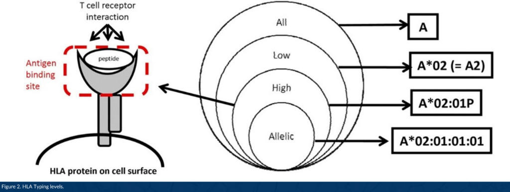
DNA-based HLA typing is often designated as low, intermediate, or high resolution. Low resolution typing corresponds to an allele group of a particular HLA gene (for example, A*01; A*02). High resolution typing corresponds to a set of alleles that encode one specific HLA protein region called the antigen binding site, used to present a peptide to immune cells (for example, A*01:02; A*02:01, Figure 2).1 High resolution typing resolves ambiguities resulting from substitutions located within exons 2 and 3 for class I HLA genes, and exon 2 for class II genes located within coding regions. This level of high resolution is usually referred to as 1x resolution. When high resolution HLA typing includes non-coding regions and allele sequences that are not expressed as cell-surface proteins, high resolution typing is termed 2x, 3x, 4x, etc.
Along with other newly discovered biomarkers, HLA phenotype and genotype may be used to predict therapeutic outcomes in a wide range of indications, including cancer. In a recent study analyzing thousands of immuno-oncology patients, researchers from the National Cancer Institute found a link between HLA-A*03 allele carriers and poor response to different checkpoint inhibitor treatments in multiple types of cancer.2 This is one of many examples of how top-quality, highly characterized cellular starting materials enable researchers with accurate donor selection and help clients move efficiently through the pre-clinical, clinical, and commercial phases of development to advance the delivery of life-saving, breakthrough therapies.
All of OrganaBio’s cell products are HLA typed by next (third) generation sequencing (NGS) of all 6 HLA genes and high resolution results are reported. High resolution genotyping by NGS is performed for HLA-A, B, C, DR, DQ, DP, and scientists can pick cell products from donors with common or rare HLA alleles. Request a copy of our inventory report here.
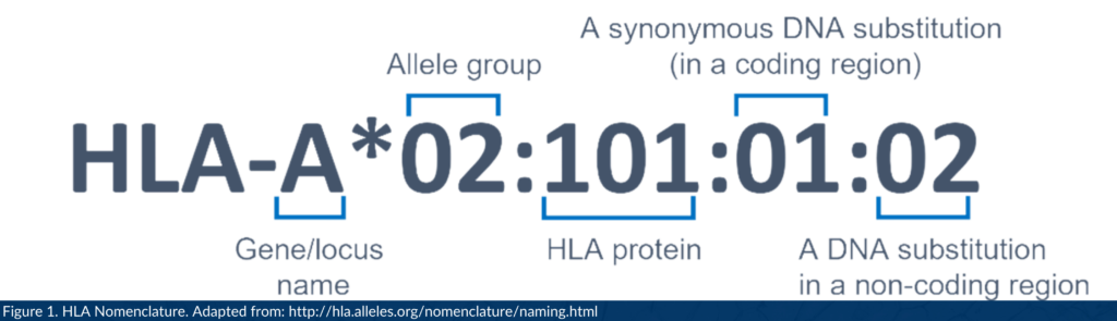
A description of the parts in an HLA name. Allele group; the allele’s antigen type, typically found by serotyping. HLA protein; the peptide for which the allele codes. Synonymous DNA substitution in a coding region; an allele variant with a different DNA sequence that produces the same protein. DNA substitution in a non-coding region; identifies a sequence polymorphism/mutation outside of the coding region.
References
- Nunes E, Heslop H, Fernandez-Vina M, Taves C, Wagenknecht DR, Eisenbrey AB, Fischer G, Poulton K, Wacker K, Hurley CK, Noreen H, Sacchi N. Definitions of histocompatibility typing terms. Blood. 2011 Dec 1;118(23):e180-3.
- Naranbhai V, Viard M, Dean M, Groha S, Braun DA, Labaki C, Shukla SA, Yuki Y, Shah P, Chin K, Wind-Rotolo M, Mu XJ, Robbins PB, Gusev A, Choueiri TK, Gulley JL, Carrington M. HLA-A*03 and response to immune checkpoint blockade in cancer: an epidemiological biomarker study. Lancet Oncol. 2022 Jan;23(1):172-184.
HLA-typed donors
Need donors matched on specific alleles?
Every donor in the OrganaBio pool is genotyped by high-resolution sequencing across HLA-A, B, C, DR, DQ and DP, with KIR genotyping available, so material can be selected by haplotype rather than screened after it arrives.
What HLA stands for, and what the letters after it mean
HLA is human leukocyte antigen. It is the human name for the major histocompatibility complex, the gene cluster on the short arm of chromosome 6 that encodes the molecules your immune system uses to tell self from non-self. Every nucleated cell in your body displays them. They are, functionally, the identity documents your cells carry.
The reason HLA is difficult is that it is the most polymorphic region in the human genome. Tens of thousands of alleles have been catalogued and the number keeps climbing. That variability is a survival advantage for the species and an engineering problem for anyone matching donor to recipient, or trying to reproduce an experiment across donors.
Reading an HLA result
A result like HLA-A*02:01:01:02 looks impenetrable until you know it is four fields separated by colons, each one narrower than the last.
| Component | Example | What it tells you |
|---|---|---|
| Gene | HLA-A | Which locus |
| Field 1, allele group | *02 | Roughly corresponds to the old serological type |
| Field 2, protein | :01 | A distinct protein sequence. This is the level most work needs. |
| Field 3, synonymous coding change | :01 | Same protein, different DNA in the coding region |
| Field 4, non-coding change | :02 | Difference outside the coding sequence |
Two fields is high resolution and is what most cell therapy and immunology work is specified against. One field is low resolution and is rarely sufficient for anything beyond a first pass. Suffixes carry meaning too: an N indicates a null allele that is not expressed, an L indicates low surface expression. A null allele reported without the suffix being read is a genuine source of downstream confusion.
Class I, class II and class III
| Class I | Class II | |
|---|---|---|
| Classical genes | HLA-A, HLA-B, HLA-C | HLA-DR, HLA-DQ, HLA-DP |
| Found on | All nucleated cells | Antigen-presenting cells: dendritic cells, macrophages, B cells |
| Presents peptide to | CD8+ cytotoxic T cells | CD4+ helper T cells |
| Peptide source | Intracellular, so viral and tumour proteins | Extracellular, taken up and processed |
| Also read by | NK cell inhibitory receptors (KIR) | Not directly |
Class III is the odd one out. It sits between the class I and class II regions on chromosome 6 and encodes complement components, tumour necrosis factor and other immune proteins, but it does not encode antigen-presenting molecules. It gets swept into HLA discussions by geography rather than by function.
The class I row worth dwelling on is the last one. Class I is not only a T cell signal. NK cells read it too, through killer immunoglobulin-like receptors, and read its absence as a reason to kill. That is why HLA and KIR genotype have to be considered together for any NK-directed programme rather than treated as separate assays.
Haplotypes, and why HLA is inherited in blocks
HLA genes sit close together on chromosome 6 and are inherited as a linked block called a haplotype. You receive one haplotype from each parent, which means a full sibling has a one in four chance of being a full match, a one in two chance of a half match, and a one in four chance of sharing neither.
Because the genes travel together, certain allele combinations occur far more often than chance would predict, and those combinations vary substantially between populations. This is the practical reason unrelated-donor matching is markedly harder for people of non-European ancestry: the registries are less densely populated with the haplotypes they carry. It is also why a donor pool that has not been characterised for ancestry is a thinner resource than its headcount suggests.
How HLA typing is actually performed
| Method | Resolution | Where it still fits |
|---|---|---|
| Serological typing | Antigen level, low | Largely historical. Underpins the old antigen nomenclature. |
| SSP and SSO (PCR-based) | Low to intermediate | Fast screening, ambiguity common |
| Sanger sequence-based typing | High, but phase ambiguity persists | Legacy workflows |
| Next-generation sequencing | High resolution, phase-resolved, multi-locus in one run | The current standard for cell therapy starting material |
NGS displaced the earlier methods for a specific reason. Older approaches frequently returned ambiguous strings, several possible allele pairs consistent with the same result, which had to be resolved with follow-up testing. Sequencing the region directly resolves phase and returns unambiguous high-resolution types across all six classical loci in a single run.
What HLA typing is used for
Transplantation
The original application. Matching at HLA-A, B, C and DRB1 remains the core of unrelated haematopoietic stem cell transplant matching, with mismatch at these loci tracking with graft-versus-host disease and graft failure. Solid organ matching weights the loci differently and tolerates more mismatch, supported by immunosuppression.
Cell and gene therapy starting material
Allogeneic programmes need to know the HLA type of their starting material for several converging reasons: predicting alloreactivity, selecting donors whose type suits an intended patient population, building HLA-defined donor panels for comparability work, and interpreting NK behaviour where class I is the ligand for inhibitory KIR. A donor whose HLA type is unknown is a donor whose immunological behaviour is unpredictable.
Disease association and drug hypersensitivity
Specific alleles associate strongly with particular conditions and with severe adverse drug reactions. HLA-B*57:01 and abacavir hypersensitivity is the textbook example, along with HLA-B*15:02 and carbamazepine reactions in certain populations. These associations are why HLA typing appears in pharmacogenomics as well as in transplant.
Immunology and vaccine research
Which peptides a person can present is determined by their HLA type, so epitope prediction, T cell assay design and vaccine response studies all depend on knowing it. Running a T cell assay on donors of unknown HLA type and then trying to explain the variance afterwards is a common and avoidable waste.
Matching, mismatching and the language around it
Matching is usually written as a fraction. An 8/8 match means HLA-A, B, C and DRB1 matched on both inherited copies. A 10/10 adds DQB1. Higher-resolution matching at more loci generally produces better transplant outcomes, though the relationship is not uniform across loci and permissive mismatches exist, particularly at DPB1.
Antigen matching and allele matching are not the same claim. Antigen-level matching compares at the lower-resolution first field. Allele-level matching compares at high resolution. Two donors described as antigen-matched can be allele-mismatched, and the difference is clinically meaningful. When a supplier says material is HLA-matched, the resolution behind that word is the question worth asking.
Sourcing HLA-typed material
For research and cell therapy development, HLA type belongs with the material rather than in a separate document requested later. OrganaBio documents high-resolution NGS typing across six loci plus KIR as part of donor characterisation across the catalogue, including fresh and cryopreserved leukopaks, cryopreserved PBMCs, peripheral blood NK cells, pan T cells and cord blood CD34+ HSCs. Donor selection can be scoped by HLA genotype where availability allows, and eligible donors can be scheduled for repeat collection, which is what makes a defined-HLA donor panel something you can return to rather than assemble once.